
Exploiting Shape Based feature for detecting nuances in vasculature
This project contains the implementation of a supervised blood vessel segmentation technique for digital fundus images using Zernike Moment-based features. The project introduces a novel approach to detect retinal vasculature with high accuracy, aiding in the diagnosis of ophthalmologic and cardiovascular disorders.
September 1, 2019
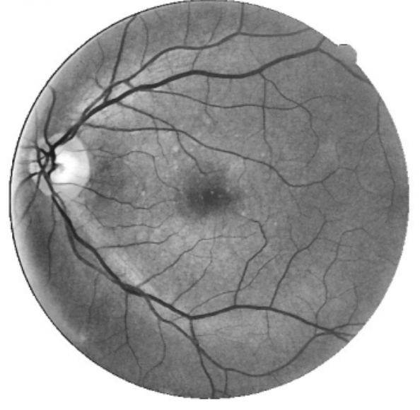
Detecting Retinal Vasculature
This repository contains the implementation of a supervised blood vessel segmentation technique for digital fundus images using Zernike Moment-based features. The project introduces a novel approach to detect retinal vasculature with high accuracy, aiding in the diagnosis of ophthalmologic and cardiovascular disorders.
Overview
Retinal vessel segmentation is crucial for diagnosing various diseases such as diabetic retinopathy, hypertension, and other cardiovascular conditions. This project combines advanced pre-processing techniques with a supervised artificial neural network (ANN) to accurately classify blood vessel pixels in fundus images.
Methodology
The segmentation pipeline consists of three main stages:

1. Preprocessing
- Input: Fundus images.
- Output: Preprocessed images with enhanced vessel structures and suppressed background noise.

Steps:
- Extract the green channel, which offers maximum contrast for vessels.
- Apply morphological operations for light reflex removal.
- Perform contrast enhancement using CLAHE.
- Generate a vessel-enhanced image via top-hat filtering.
2. Feature Extraction
- Gray-Level Features:
- Intensity differences within local windows (minimum, maximum, mean, and standard deviation).
- Zernike Moment Features:
- 36 Zernike coefficients calculated from 17×17 windows.
- Dominant coefficients selected based on maximum discriminability.


3. Classification
- Features are passed to an ANN:
- Input Layer: 11 nodes (features).
- Hidden Layers: 3 layers with 23 nodes each (tanh activation).
- Output Layer: 2 nodes (softmax for vessel/background classification).

Performance
MetricDRIVE DatasetSTARE DatasetAccuracy94.5%94.86%Sensitivity69.94%62.98%Specificity98.11%98.39%AUC93.94%95.07%
The proposed model achieves high accuracy while preserving thin blood vessels, a challenge for traditional methods.
Dataset
The method is evaluated on two publicly available datasets:
Installation
Clone the repository and install the dependencies:
git clone https://github.com/TakedoNick/Detecting_Retinal_Vasculature.git
cd Detecting_Retinal_Vasculature
pip install -r requirements.txt
Usage
Training
python train.py --data_dir <path_to_training_data>

Testing
python test.py --data_dir <path_to_testing_data> --output_dir <output_path>

Visualization
python visualize_results.py --results_dir <path_to_results>


References
References • Adapa D., Joseph Raj A. N., et al. “A supervised blood vessel segmentation technique for digital fundus images using Zernike Moment-based features.” PLOS ONE, 2020.
• DRIVE Dataset
• STARE Dataset